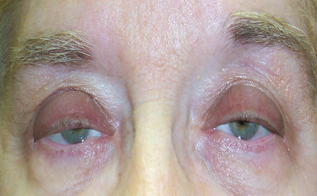
- What is ptosis?
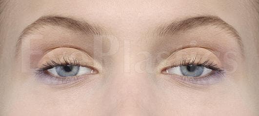
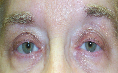
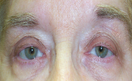
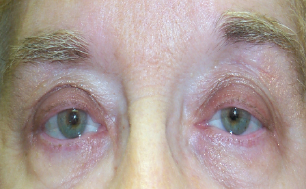
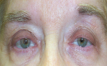
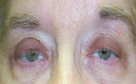
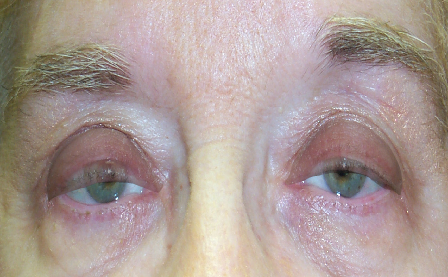
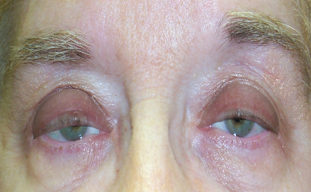
- Ptosis (TOE-sis) is drooping of the upper eyelid caused by weakness or dysfunction of the levator muscle — the muscle responsible for lifting the upper eyelid. It can affect one or both eyes and may be present from birth (congenital) or develop over time (acquired).
- What is the difference between ptosis and blepharoplasty?
- Ptosis is caused by levator muscle weakness and requires surgical repair of the muscle itself. Blepharoplasty addresses excess skin overlying the eyelid. Both conditions can cause drooping or hooding, and they often occur together. Only an oculoplastic surgeon can reliably distinguish them and perform the correct procedure.
- How is ptosis repaired surgically?
- The most common technique is levator advancement — tightening the levator aponeurosis through an external incision in the eyelid crease. If levator function is poor (as in severe congenital ptosis), a frontalis sling procedure connects the eyelid to the brow muscle. Mild ptosis in patients who respond to phenylephrine drops can be corrected with a Müller's muscle-conjunctival resection (MMCR).
- Is ptosis surgery covered by insurance?
- Yes — ptosis repair is typically covered by health insurance when the drooping eyelid causes functional visual field obstruction, documented by a formal visual field test with the eyelid in its resting position.
- What should I expect during a ptosis consultation?
- During your consultation, Dr. Fountain will perform a comprehensive eye examination, including measuring your eyelid height and assessing how well your levator muscle functions. You'll discuss your symptoms, review your medical history, and examine photographs to determine if you're a good candidate for surgery. The surgeon will explain the appropriate surgical technique for your specific condition and answer any questions about risks, recovery, and expected outcomes.
- What are the potential risks and complications of ptosis surgery?
- While ptosis repair is generally safe, potential complications include infection, bleeding, scarring, and asymmetry between the two eyelids. Some patients may experience temporary dry eyes, difficulty closing the eyelid completely, or under- or over-correction requiring revision surgery. These complications are uncommon, especially when performed by a fellowship-trained oculoplastic surgeon, and most resolve with appropriate care.
- What is the recovery timeline after ptosis surgery?
- Most patients can return to light activities within one to two weeks, though complete healing typically takes four to six weeks. During the first few days after surgery, you may experience swelling, bruising, and mild discomfort managed with prescribed medications and cold compresses. You'll have follow-up appointments to monitor healing, and Dr. Fountain will provide specific instructions about activity restrictions, eye care, and when you can resume normal routines.
- What causes a droopy eyelid (ptosis)?
- The most common cause in adults is aponeurotic ptosis, where the levator tendon stretches or detaches with age, contact-lens wear, or after eye surgery. Other causes include congenital (present from birth), neurogenic (nerve problems such as Horner's syndrome, third-nerve palsy, or myasthenia gravis), and myogenic muscle disease.
- Can ptosis be fixed without surgery?
- Mild ptosis can be improved with oxymetazoline 0.1% (Upneeq) eyedrops, which stimulate Muller's muscle to lift the lid a millimeter or two. Drops are temporary and work best for mild cases; moderate to severe ptosis is corrected surgically.
- Is ptosis repair covered by insurance?
- Ptosis repair is often covered when it is functional -- when a formal visual-field test documents that the lid obstructs your superior vision. Photographs and prior authorization are usually required. If a cosmetic blepharoplasty is done at the same time, the skin portion is billed separately as cosmetic.
- How is ptosis diagnosed?
- An oculoplastic surgeon measures margin reflex distance (MRD-1, normally about 4-5 mm; ptosis at 2 mm or less) and levator function (good is 10 mm or more, poor is 4 mm or less), and may use a phenylephrine test and visual-field testing. These measurements determine which repair is appropriate.