Cosmetic
Autologous fat transfer to restore facial volume — a natural, long-lasting alternative to dermal fillers for periorbital and midface rejuvenation.
Medically reviewed by Tamara R. Fountain, MDOculoplastic SurgeonLast updated June 2026
Facial fat grafting — also called autologous fat transfer, lipofilling, or fat injection — is the surgical relocation of a patient’s own fat from areas of relative excess (typically the abdomen or inner thigh) to areas of the face that have lost volume with age. Unlike synthetic dermal fillers, which are temporary placeholders, transplanted fat can become permanently incorporated into the recipient tissue, bringing along its own blood supply and a population of adipose-derived stem cells thought to improve overlying skin quality.
For the periorbital region — the temples, brows, upper eyelid sulcus, tear troughs, and cheeks — fat grafting addresses a specific aging problem that lifting alone cannot fix: volume loss. As we age, fat compartments around the eye atrophy, the bony orbit expands, and the skin retracts inward, producing a hollow, skeletonized appearance even in patients with otherwise healthy tissue. Repositioning lax skin without restoring this lost volume often produces an over-pulled, unnatural result. Fat grafting restores the youthful contour from underneath.
Oculoplastic surgeons are uniquely qualified to perform periorbital fat grafting because they understand the delicate anatomy of the eyelid and orbit at a level few other specialists do. The margin for error is measured in tenths of a milliliter, and the consequences of poor technique — lumpiness, overcorrection, or, in rare cases, vascular complications — are highly visible and difficult to reverse.
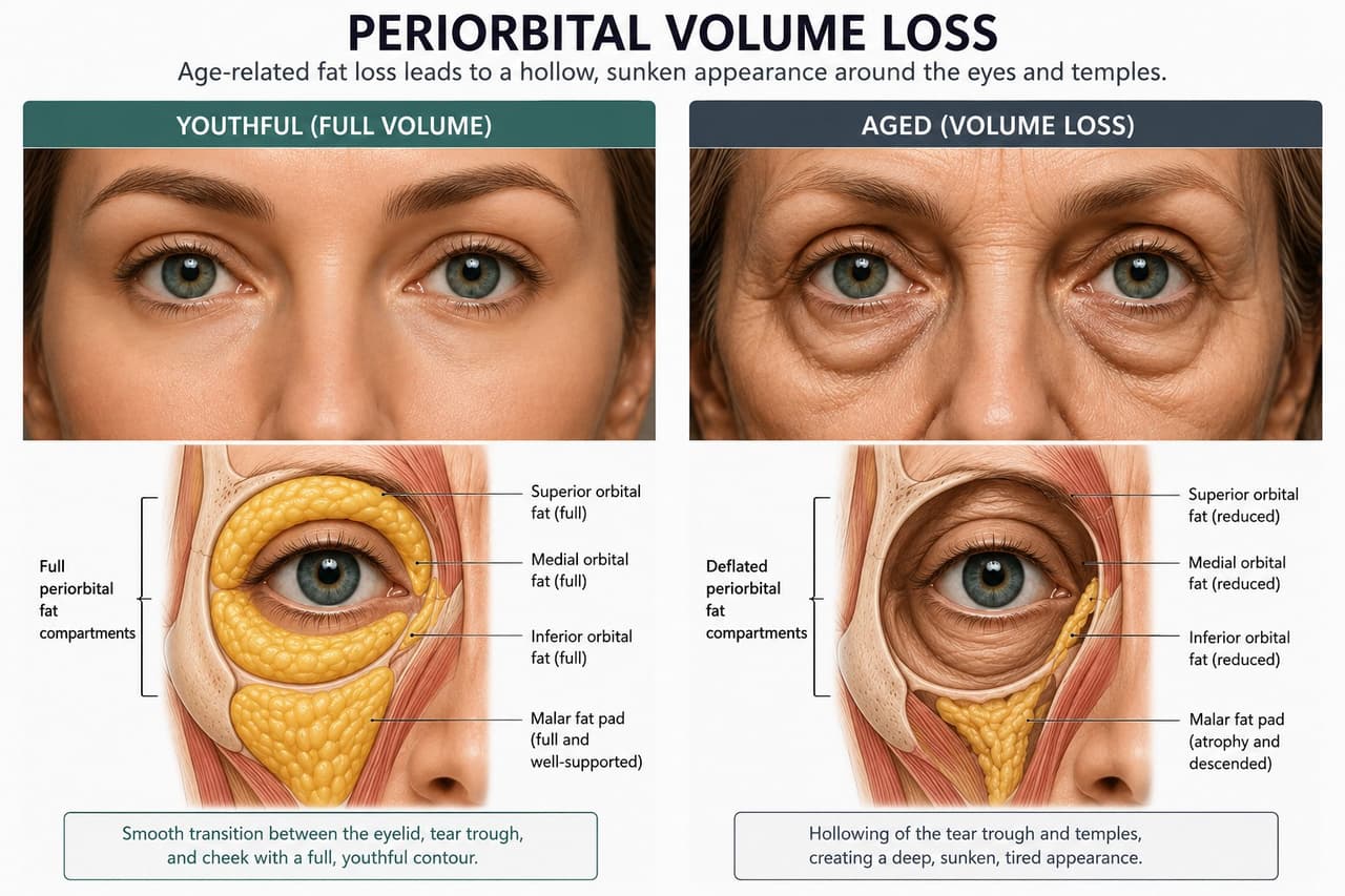
The periorbital region encompasses several distinct zones, each requiring a tailored grafting strategy. These areas are particularly demanding because the skin is the thinnest on the body, the underlying tissue is sparse, and even small irregularities are immediately visible.
Temporal hollowing creates a gaunt, ill appearance and causes the lateral brow to descend. Restoring temple volume lifts the brow tail indirectly and broadens the upper face, often producing one of the most rejuvenating effects of any single intervention. Temple grafting is typically performed in the deep plane just above periosteum, both to stay clear of the superficial temporal vessels (a recognized vascular danger zone) and to avoid visible contour irregularities.
The hollow upper sulcus — often called the “A-frame deformity” — is a hallmark of aging and is also commonly seen after aggressive upper blepharoplasty performed decades earlier. Small-volume grafting (often 1–2 mL per side) into the sub-brow fat pad and pre-septal plane can restore the youthful convexity without weighing down the lid.
The tear trough is perhaps the most technically challenging area on the face for any volumizing procedure. Fat must be placed deep, against bone, beneath the orbicularis muscle, in micro-aliquots. Superficial placement produces visible lumps, yellow discoloration, or the dreaded chronic puffiness that can persist for years.
Patients considering tear trough treatment should review the differences between fat grafting, hyaluronic acid Fillers, and surgical Tear Trough correction with their surgeon — each has distinct advantages depending on anatomy.
Volumizing the malar eminence and anterior cheek supports the lower lid from below, softens the nasolabial fold, and re-creates the youthful “ogee curve” in profile. Midface fat grafting is often combined with a Midface Lift or lower blepharoplasty for comprehensive rejuvenation.
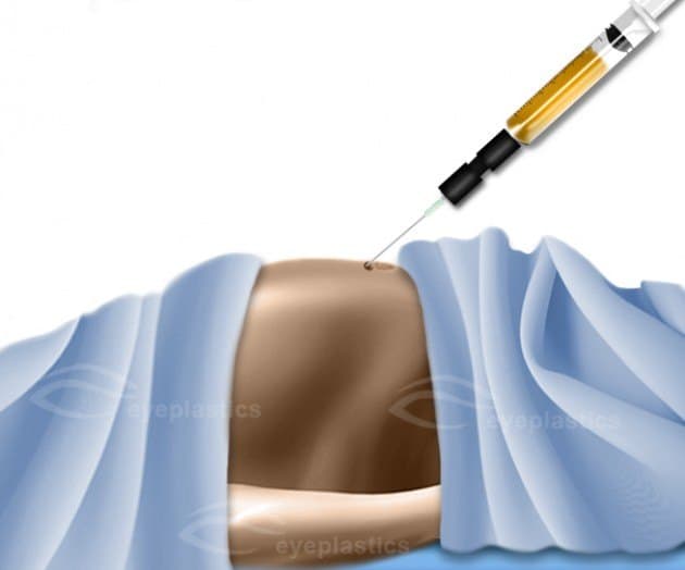
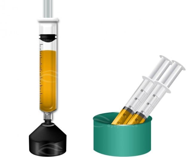
The quality of the final result depends as much on how the fat is harvested as on how it is injected. Fat is a living tissue; rough handling, exposure to air, or excessive negative pressure during harvest will kill adipocytes and reduce graft survival.
Common donor sites in order of preference for facial grafting:
For periorbital grafting, only 10–30 mL of harvested fat is typically needed, making donor site morbidity minimal. The harvest is performed with tumescent local anesthesia using a small (2–3 mm) blunt cannula and low-vacuum aspiration — either gentle syringe suction or a dedicated low-pressure pump.
Once harvested, the lipoaspirate contains a mixture of intact adipocytes, ruptured cells releasing free oil, blood, tumescent fluid, and local anesthetic. All of these contaminants must be removed before the graft is injected, or they will provoke inflammation and reduce viability.
Three processing techniques dominate current practice:
Many oculoplastic surgeons further refine the graft for periorbital use through micro-fat or nano-fat processing — passing the lipoaspirate through progressively smaller filters or inter-syringe transfers to produce particles small enough to inject through a 27-gauge cannula into the eyelid skin without lumps.

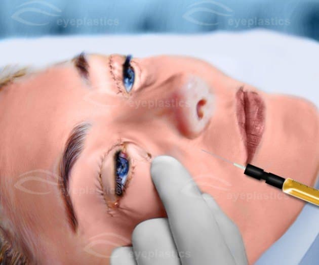
Injection technique is where periorbital fat grafting succeeds or fails. The fundamental principles, articulated by Sydney Coleman and refined by subsequent surgeons, remain unchanged:
Important: Vascular embolism from fat injection — though rare — is among the most feared complications in aesthetic surgery and has caused blindness and stroke when injection is performed with sharp needles in the glabella, tear trough, or temple. The use of blunt cannulas with low injection pressure is non-negotiable in the periorbital region.
One of the most common questions patients ask is how fat grafting compares to hyaluronic acid fillers. Both restore volume, but they differ in nearly every other respect.
| Feature | Fat Grafting | HA Fillers |
|---|---|---|
| Longevity | Permanent for surviving cells (typically 40–70% take) | 6–18 months depending on product and area |
| Setting | Operating room, sedation or general | Office, topical anesthetic |
| Recovery | 1–2 weeks of bruising & swelling | Hours to days |
| Reversibility | Not reversible; revision requires surgery | Dissolvable with hyaluronidase |
| Skin quality benefit | Yes — stem cell effect on overlying skin | Minimal |
| Volume capacity | Large (10s of mL) | Smaller, cost-limited |
| Predictability | Variable take rate | Highly predictable |
In practice, fat grafting is the right choice for patients with significant global volume loss who want a one-time, long-lasting solution and are willing to accept surgical downtime. Fillers remain superior for first-time volume patients, precise spot corrections, and anyone who wants reversibility.
Fat grafting is rarely performed in isolation. Most oculoplastic surgeons integrate it into a comprehensive rejuvenation plan because volume restoration amplifies the results of every other procedure.
Common combinations:
Patients undergoing combined procedures benefit from a single recovery period and from the synergistic effect of treating both skin laxity and volume loss. Learn more about Blepharoplasty and Midface Lift for complementary options.
Although fat grafting uses the patient’s own tissue and avoids the risk of allergic reaction, it carries its own specific set of risks that every patient should understand.
Important: Vascular embolism — inadvertent injection of fat into a facial artery — can cause skin necrosis, blindness, or stroke. This catastrophic complication is the reason periorbital fat grafting should only be performed by surgeons with detailed knowledge of facial vascular anatomy using blunt cannulas, slow injection, and low pressure.
Recovery from periorbital fat grafting is shaped primarily by the swelling and bruising response, which is more pronounced than with filler treatment because of the additional trauma of cannula passes through tissue.
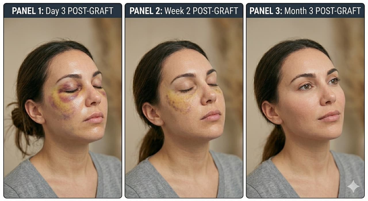
Significant swelling and bruising are expected, particularly around the eyes if periorbital grafting was performed. The face will look noticeably overcorrected — this is normal and reflects edema rather than the final result. Cold compresses (gentle, not applied with pressure) for the first 48 hours help limit swelling. Patients sleep with the head elevated and avoid bending, lifting, or strenuous activity.
Bruising resolves and most visible swelling subsides. The face still appears fuller than the final result. Patients can typically return to work within 7–10 days, sometimes longer if extensive grafting was performed. Makeup can be used to camouflage residual discoloration after sutures (if any) are removed.
The grafted fat undergoes its critical period of revascularization. Fat that successfully connects to new blood vessels survives; the rest gradually reabsorbs. The final volume becomes apparent during this window. Patients should avoid significant weight loss during recovery, as grafted fat cells respond to overall body weight changes — substantial weight loss can shrink the graft.
Final results stabilize. At this point, the surgeon and patient can assess whether touch-up grafting is desired. Roughly 20–30% of patients elect a small secondary procedure to refine areas of under-correction; this is planned for from the outset and is part of the natural arc of fat grafting.
Surviving grafted fat behaves like the donor-site tissue from which it came — meaning it can grow with weight gain and shrink with weight loss. The graft itself does not “age” faster than native tissue, and many patients enjoy stable volume restoration for a decade or more. Continued natural facial aging proceeds normally elsewhere, which is why fat grafting is often repeated in modest amounts every 5–10 years for ongoing maintenance.
Periorbital fat grafting is a deceptively difficult procedure. The technical steps are simple to describe but require years of refinement to execute well. An ASOPRS fellowship-trained oculoplastic surgeon brings unmatched understanding of eyelid and orbital anatomy, vascular safety, and the aesthetic principles that distinguish a natural result from a surgical one. If you are considering fat grafting around the eyes — whether alone or in combination with blepharoplasty or brow surgery — we encourage you to Find a Doctor in your area with the specialized training to perform this procedure safely and beautifully.
Your Surgeon
Tamara R. Fountain, MD
Ophthalmology Partners, Ltd.
🏅 ASOPRS Fellowship Trained
Schedule a consultation with Tamara R. Fountain, MD to learn if this procedure is right for you.